Atrial Septal Defect in Canine
Medical History
Breed: Welsh Corgi
Age: 5 months
Gender: Male, intact
Body Weight: 4.2 kg
The dog was initially noted to have a mild heart murmur during a routine health examination at 3 months of age, with no obvious clinical abnormalities at that time.
In recent weeks, however, the owner reported that the 5-month-old Corgi became easily fatigued during play, showed occasional dry coughing, and demonstrated reduced exercise tolerance compared to before. Mildly increased respiratory effort and louder breathing sounds were also observed during rest and sleep.
Ultrasound Findings
These findings supported the diagnosis of atrial septal defect.
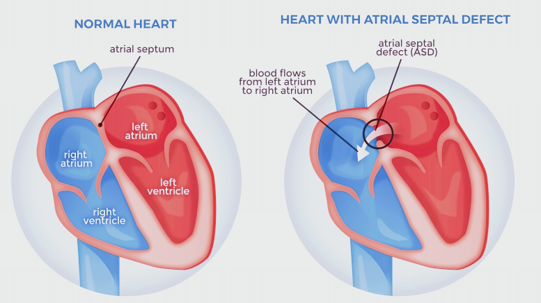
What Is an ASD?
An Atrial Septal Defect (ASD) is a congenital heart abnormality in which the wall separating the left and right atria fails to close completely during fetal development. As a result, blood can pass abnormally between the two atrial chambers.
What Causes ASD?
ASD develops during embryonic heart formation and is present at birth.It is not caused by diet, environment, or management after birth, and in most cases, the exact cause remains unknown.
How Does ASD Affect the Heart?
Because pressure in the left atrium is normally higher than in the right atrium, blood flows from left to right through the defect. Over time, this abnormal shunting can lead to:
Increased blood volume in the right heart
Enlargement of the right atrium and right ventricle
Increased blood flow to the lungs
If left untreated, these changes may progress and affect overall cardiac function.
Type of ASD?
Ostium Secundum ASD (Most Common Type): A defect in the middle of the atrial septum (fossa ovalis), often small and well tolerated.
Ostium Primum ASD: A defect in the lower atrial septum, often associated with other defects like atrioventricular valve abnormalities. More severe than secundum ASDs.
Sinus Venosus ASD: A defect near the entry of the vena cava, sometimes affecting pulmonary vein connections. Often requires surgical correction.
Which Dogs Are at Risk?
ASD can occur in dogs of any breed.It is most often detected in young dogs but may also be discovered incidentally in adults during cardiac screening. Some cases remain asymptomatic for years, especially when the defect is small.
Why Is It Dangerous?
Although some atrial septal defects are mild, larger or long-standing defects may result in:
Chronic right heart volume overload
Pulmonary vascular remodeling
Secondary pulmonary hypertension
Right-sided heart failure in advanced cases
Early recognition is essential to prevent long-term complications.
Echocardiography is the most important and reliable diagnostic tool for atrial septal defect in dogs.
Ultrasound allows clinicians to:
Directly visualize the atrial septum
Identify the size and location of the defect
Assess shunt direction using color Doppler
Evaluate secondary changes in cardiac chambers
Monitor pulmonary circulation and disease progression
Without ultrasound, ASD may be easily overlooked, especially in dogs with mild or nonspecific clinical signs.
The final diagnosis of atrial septal defect is based on echocardiographic confirmation of the septal defect and abnormal atrial blood flow.
Clinical management depends on the size of the defect, the degree of shunting, and the presence of secondary cardiac or pulmonary changes.
Regular follow-up with cardiac ultrasound is recommended to monitor disease progression and guide treatment decisions.
Canine atrial septal defect is a congenital heart disease that may remain silent in early stages but can lead to significant cardiac consequences over time.
Comprehensive echocardiographic evaluation plays a critical role in early detection, accurate diagnosis, and long-term management.
By identifying structural defects and their hemodynamic impact, ultrasound enables clinicians to make informed decisions and optimize outcomes for affected dogs.