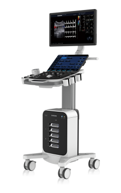
SonoMax VET
Premium Imaging for Animal Healthcare
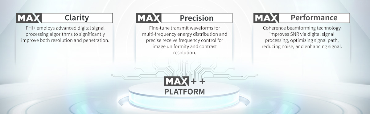
Pioneering MAX++ platform with insight.
Elevate your imaging with the most advanced ultrasound technology.
Effortless elegance, intuitive workflow, and exceptional performance.
Specifications
Latest MAX++ Platform
23.8 Large Size HD LED Screen
15.6" Angle 30° Adjustable Touch Screen
5 Transducer Connectors with Stylish Lights
25cm Flexible Height Adjustment
Smaller Projected Area
Brand-new Keyboard for Concise Workflow
Fast and Reliable After-sales Service
Application
The revolutionary cart-based ultrasound CHISON SonoMax Vet is designed to elevate veterinary imaging to new heights. With a sleek design, intelligent features, exceptional performance, and ergonomic adaptability, SonoMax Vet redefines what’s possible in veterinary ultrasound, setting a new standard for the future of animal care.
· Larger screen for enhanced clarity, with 5 transducer connectors featuring stylish lights—combining practicality with modern aesthetics.
· Exceptional performance powered by the MAX++ platform, tailored for versatile veterinary applications, from small animals to equine and farm animals.
· Simplify workflows and maximize efficiency, enabling veterinarians to focus on delivering precise and compassionate care to every patient.
Features

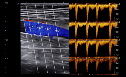
SonoPW
The PW sample gate can be set up to 4.
Synchronous display of image and calculation results.
Clinical applications: mainly used for measuring bloodflow velocity before and after arterial stenosis in animalsand for assessing the degree of embolism.

TDI-SonoPW
Myocardial movements were evaluated simultaneously.
Up to 4 sample gates.
Provides more diagnostic information.
Offers a more effective diagnostic basis.
Clinical applications: Observing the different movementspeeds of the heart can determine whether there arelocal lesions and can also evaluate early diastolic function.

Strain and Strain Rate
It is a tissue Doppler ultrasound technique used for evaluating myocardial function.
Ventricular diastolic function and wallmotion can be quantitatively evaluated.

Elastography
Utilizes the tissue's own pulsation to generatepressure and form an elastic image.
Equipped with quantitative methods (gray scaleto elastic area ratio, strain rate ratio).
Clinical application: Assessment of lesions insmall organs and superficial tissues, such asthe animal liver.
- Demo
- Transducers/Accessory


Related Products

Leave Us A Message