SonoPort VET
Premium Imaging for Animal Healthcare
Physics-Defying Portability
AI-Powered Intelligence
Premium Diagnostic Confidence
Anywhere Care Happens
Specifications
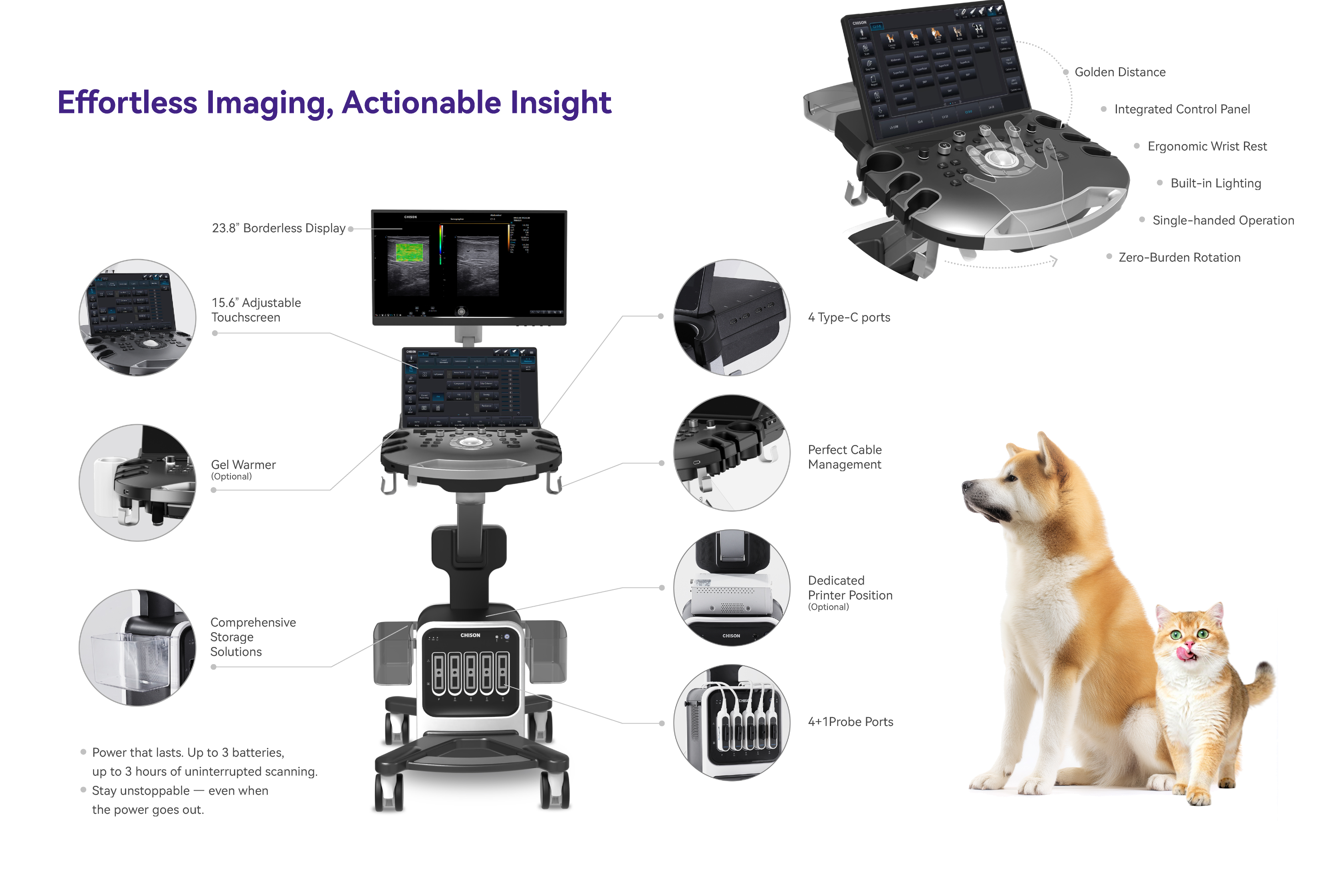
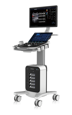
SonoPort 8 VET defies convention with physics-defying portability and Al intelligencebuilt for the unique needs of animal anatomy. This empowered diagnostic platformbrings the full power of precision imaging out of the static room and directly to thepoint of care-whether in the clinic, barn, or field-delivering insights instantly, wher-ever they are needed.
Application
Features

SonoPW
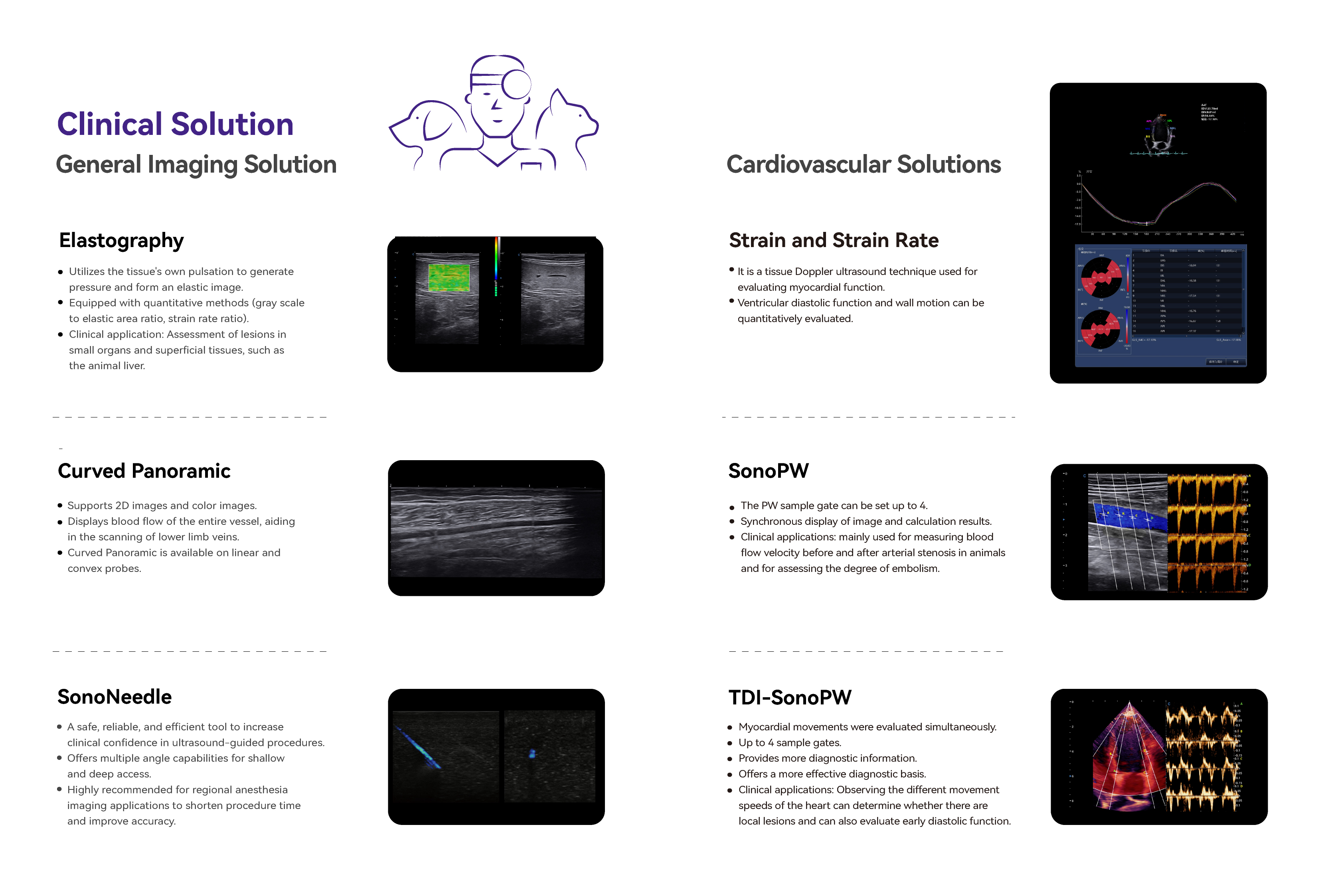
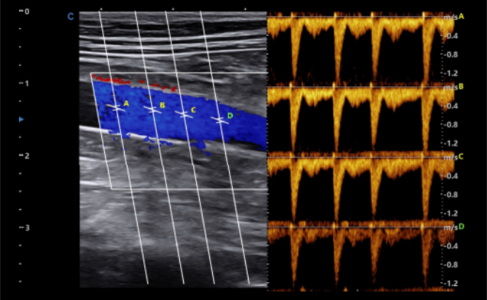
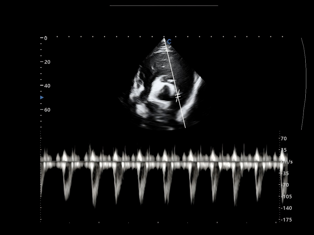
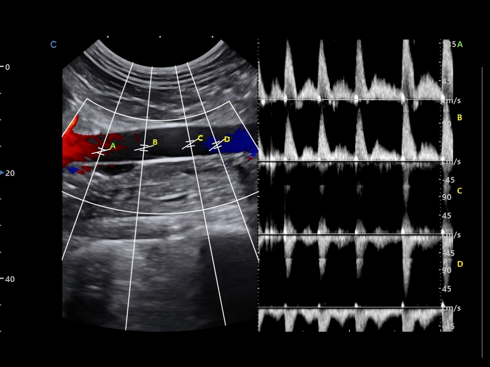
The PW sample gate can be set up to 4.
Synchronous display of image and calculation results.
Clinical applications: mainly used for measuring bloodflow velocity before and after arterial stenosis in animalsand for assessing the degree of embolism.
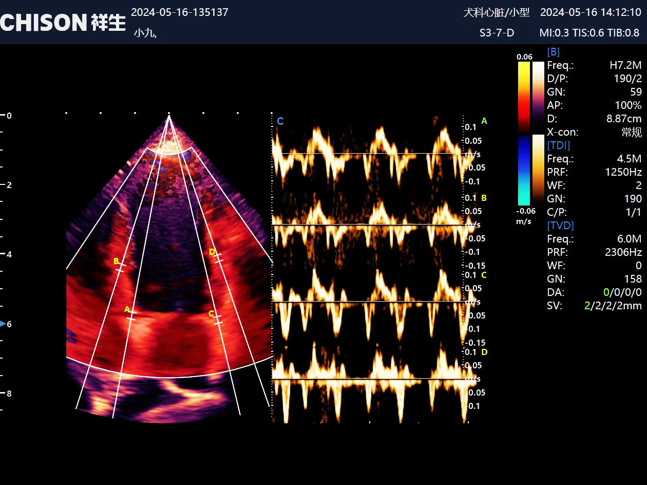
TDI-SonoPW
Myocardial movements were evaluated simultaneously.
Up to 4 sample gates.
Provides more diagnostic information.
Offers a more effective diagnostic basis.
Clinical applications: Observing the different movementspeeds of the heart can determine whether there arelocal lesions and can also evaluate early diastolic function.
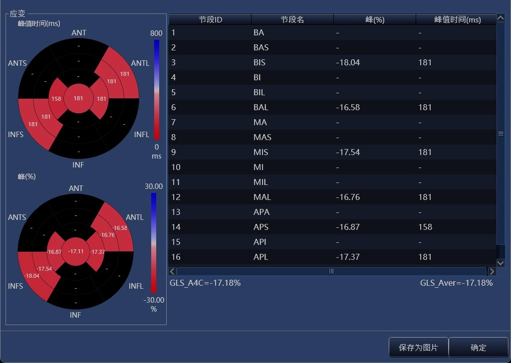
Strain and Strain Rate
It is a tissue Doppler ultrasound technique used for evaluating myocardial function.
Ventricular diastolic function and wallmotion can be quantitatively evaluated.
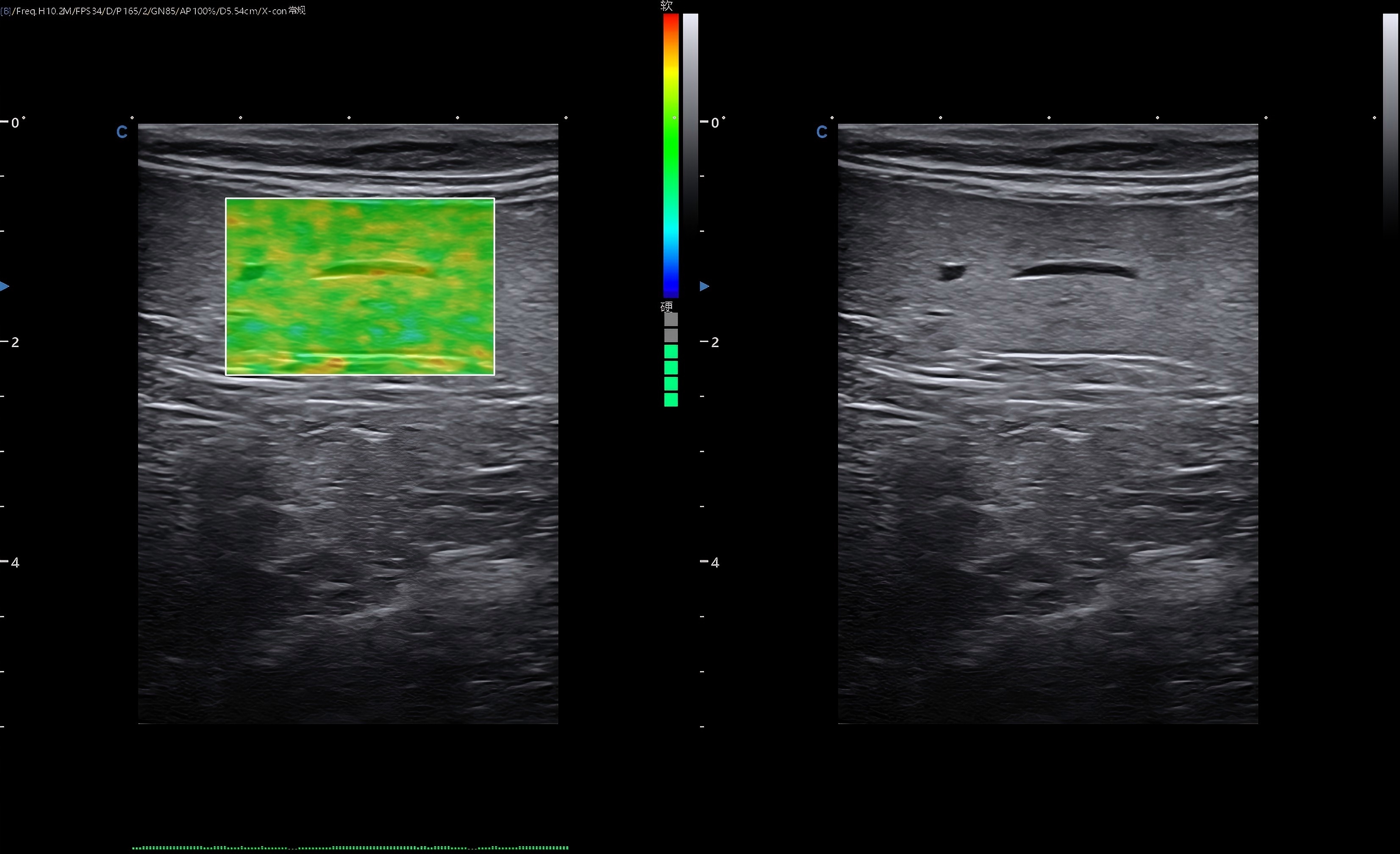
Elastography
Utilizes the tissue's own pulsation to generatepressure and form an elastic image.
Equipped with quantitative methods (gray scaleto elastic area ratio, strain rate ratio).
Clinical application: Assessment of lesions insmall organs and superficial tissues, such asthe animal liver.
- Demo
- Transducers/Accessory
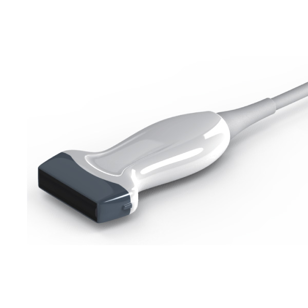
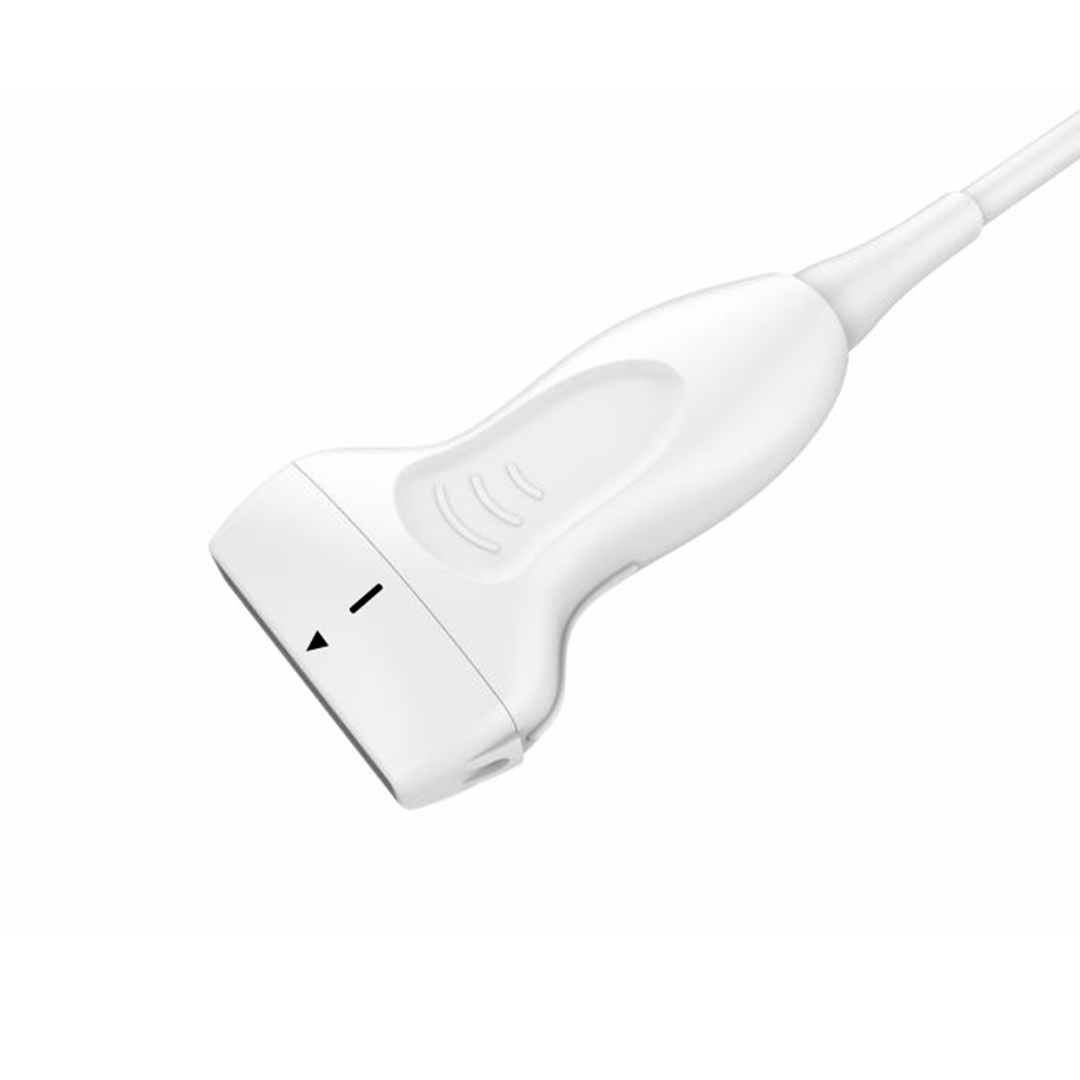
Related Products

Leave Us A Message