How to Get a Reliable Cardiac View in Small Animals
2026-05-13
In cardiac ultrasound, we often focus on measurements and interpretation.
But in practice, most problems start much earlier.
Not with the machine.
Not with the settings.
But with something far more basic:
"How the patient is positioned — and how the probe is oriented."
Because before any measurement can be trusted,
the image itself must be correct.
1. Positioning-Creating Access to the Heart
A reliable cardiac scan always begins with proper positioning.
In small animal echocardiography, two standard positions are used:
Right lateral recumbency
→ for right parasternal views
→ long-axis and short-axis imaging
Left lateral recumbency
→ for left apical views
→ four-chamber, five-chamber, Doppler evaluation
" Standard echocardiographic windows are accessed through proper lateral positioning of the patient."
But in clinical practice, correct positioning is not just about left or right.
It comes down to three essentials:
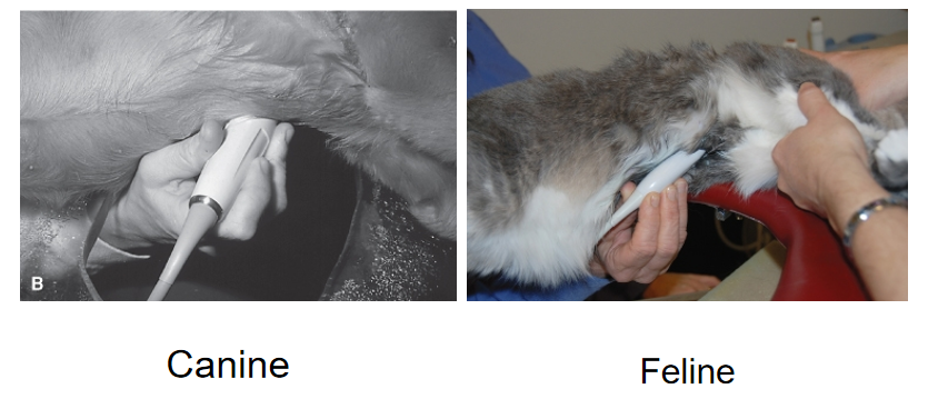
Stability — the patient remains as still as possible
Access — the probe can reach the cardiac window (behind the elbow)
Alignment — the thorax is not twisted or compressed
Even small deviations can lead to:
Distorted cardiac structures
Poor image quality
Inconsistent measurements
Positioning determines whether you can see the heart.
Once the patient is positioned correctly, the next step is orientation.
And this is where many beginners struggle.
Because getting “a heart image” is not enough.
The image must be obtained from a standard anatomical plane.
This depends on one critical element:
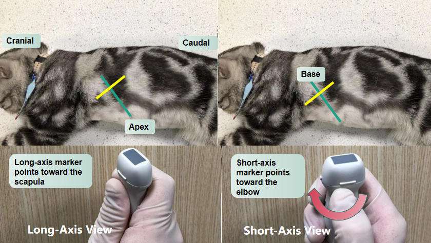
The probe marker.
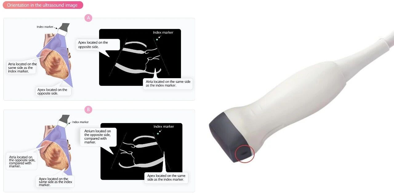
The probe marker defines the direction of the image on the screen.
If the marker is inconsistent:
The same structure may appear reversed
Standard views become difficult to reproduce
Measurements lose comparability
"Consistent probe orientation is essential for obtaining standardized echocardiographic views and accurate measurements."
A practical way to understand orientation is this:
Positioning allows access to the heart
Orientation defines how the heart is displayed
Both are required.
Without positioning → you may not see the heart clearly
Without orientation → what you see may not be interpretable
In real clinical settings, perfect conditions are rare.
Animals move.
Probe angles shift.
Views are adjusted constantly.
So the goal is not perfection.
The goal is consistency.
Use the same positioning approach
Keep probe orientation consistent
Adjust gradually, not randomly
Because in echocardiography, small variations can lead to significant differences.
"Variability in acquisition technique can affect the reproducibility of echocardiographic measurements."
"
If positioning allows access,
and orientation defines the view,
then the next step becomes clear:
How do we consistently obtain standard cardiac views?
References
[1] Thomas, W. P., et al.
Diagnostic Atlas of Canine and Feline Echocardiography. Elsevier.